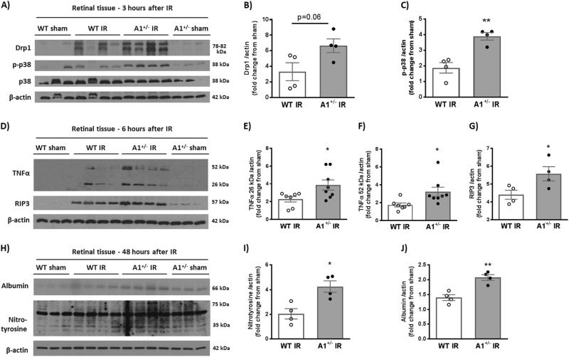
Fig. 3. A1 deletion increases inflammation, oxidative stress, and necroptosis markers after IR injury.
a Western blotting on retinal tissues collected at 3 h after IR showed higher levels of the stress marker p-p38 in A1+/− mice compared to WT after IR injury. There was also a trend towards higher levels of the mitochondrial fission protein, Drp1. b, c show quantification of Drp1 and p-p38 respectively. d Analysis at 6 h after IR injury showed a similar trend with increased TNF-α (26 kDa, membrane bound and 52 kDa, homotrimeric form), and RIP3 in A1+/− retinas as compared to WT. e−g show quantification of TNF-α bands and RIP3 respectively. h A1+/− mice showed increased nitrotyrosine (marker for peroxynitrite-mediated oxidative stress via protein nitration) and albumin extravasation (measure of permeability) at 48 h after IR injury. i, j show quantification of nitrotyrosine and albumin western blotting respectively. *p < 0.05, **p < 0.01

