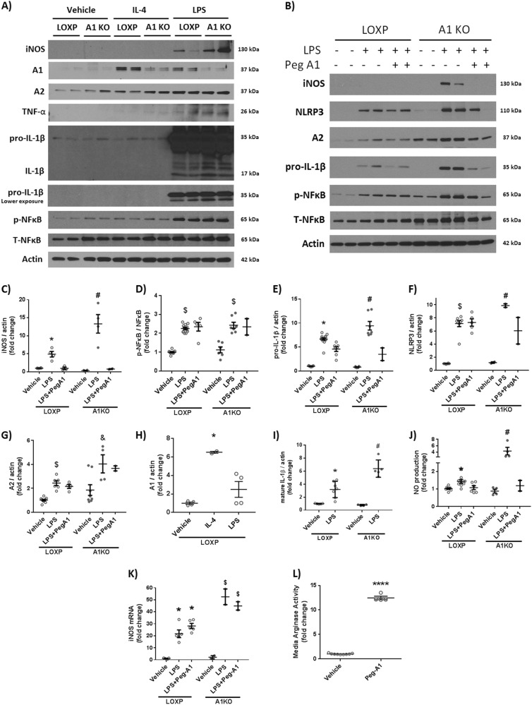
Fig. 6. Macrophages lacking A1 show a more pronounced inflammatory response to LPS stimulation in vitro and PEG-A1 treatment mitigates it.
a Western blotting of peritoneal macrophage cell lysates showed increased iNOS expression, TNF-α, and pro-IL-1β upon LPS stimulation which was further augmented in A1 KO macrophages. b PEG-A1 treatment (1 μg/ml) reduced this inflammatory response. c−i Quantification of western blot bands. *p < 0.05 vs. loxP vehicle and loxP LPS + PEG-A1, #p < 0.05 vs. loxP LPS, A1KO vehicle and A1KO LPS + PEG-A1, $p < 0.05 vs. respective vehicle. &p < 0.05 vs. loxP LPS. j A1 KO macrophages showed more nitric oxide (NO) release into the media in response to LPS, as measured using NO analyzer and this was ameliorated by PEG-A1, *p < 0.05 vs. vehicle loxP, #p < 0.05 vs. loxP LPS, A1KO vehicle, A1KO LPS + PEG-A1. k RT-PCR on BMDMs showed increased iNOS mRNA expression with LPS that was further increased in A1 KO macrophages. PEG-A1 treatment did not affect iNOS mRNA expression *p < 0.05 vs. loxP vehicle, $p < 0.05 vs. respective loxP group. l Media from wells treated with PEG-A1 show marked elevation of arginase activity (12-fold increase compared to control) at the end of a 24 h incubation, ****p < 0.0001

