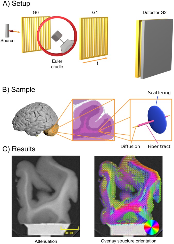
Figure 1.
A general setup overview is shown in (A). A standard X-ray imaging setup with a source and a detector is augmented with two absorption gratings G0 and G2 and one phase grating G1. Additionally, a Euler cradle is used to rotate the sample in a fully three-dimensional manner. Additionally, the grating orientation t and one X-ray direction l are illustrated. In B) we illustrate the location of the cerebellum within the human brain (left)32. In the middle of (B) we show a schematic histology image of the cerebellum with H&E stain. The center region of the histology image shows the fiber tracts located in the white matter of human brain tissue, aligning with it. Finally, on the right side of (C) we sketch the relationship of the fiber tracts, the diffusion MRI and the assumed scattering signal. In (C) we show a slice of conventional attenuation based CT on the left, and on the right an overlay of the additional directional information obtained by the proposed method in this paper.

