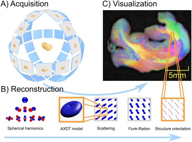
Figure 2.
In (A)32 we illustrate the acquisition scheme, which in contrast to standard CT, requires fully three-dimensional rotation of the sample instead of one single axis of rotation. Subfigure (B) illustrates the reconstruction pipeline. Firstly, the scattering in each position of the specimen is modeled via spherical harmonics. Secondly, this scattering is reconstructed using tomographic reconstruction. Thirdly, the scattering information is transformed via the Funk-Radon and local maxima are extracted to obtain the orientations of the scattering profiles. In (C) we sketch the embedding of these reconstructed fiber orientation in our final result.

