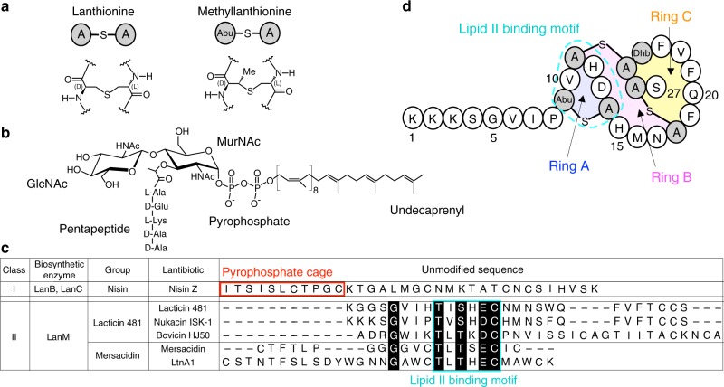
Fig. 1.
Primary structure of nukacin ISK-1 and lipid II. a Chemical structure of lanthionine and methyllanthione. b Chemical structure of lipid II. GlcNAc, N-acetylglucosamine; MurNAc, N-acetylmuramic acid. c Sequence alignment of the unmodified sequences of the class I and II lantibiotics. Highly conserved residues are highlighted in black. The lipid II pyrophosphate binding cage of the class I lantibiotics are enclosed by red lines. The lipid II binding motifs of the class II lantibiotics are enclosed by cyan lines. d Primary structure of nukacin ISK-1. Special regions are shaded and labeled: Ring A region (Abu9-Ala14), Ring B region (His15-Ala18), and Ring C region (Phe19-Ala26). Unusual amino acid residues are shown by shaded circles: Abu represents α-amino butyric acid; Dhb, dehydrobutyrine; Abu-S-A, methyllanthionine, and A-S-A, lanthionine, where “-S-“ denotes a monosulfide linkage

