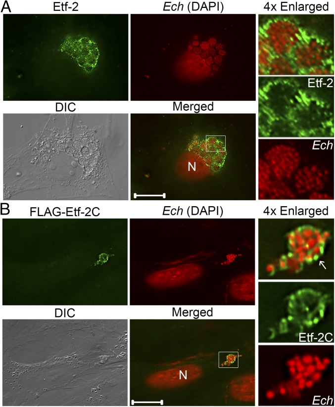
Fig. 1.
Native and FLAG-Etf-2 cloned into the Ehrlichia genome are secreted into host-cell cytoplasm and localize to the ehrlichial inclusion membrane. (A) Etf-2 was secreted and subsequently was trafficked to Ehrlichia-containing inclusion membranes. E. chaffeensis (Ech)-infected RF/6A cells at 2 dpi were fixed and labeled with rabbit anti–Etf-2C IgG and AF488 anti-rabbit IgG. (Scale bar: 10 μm.) (B) FLAG-Etf-2C cloned and expressed in Ehrlichia was secreted, and localized on inclusion membranes (white arrow). Transformed E. chaffeensis expressing FLAG-Etf-2C were selected with antibiotics in DH82 cells and used to infect RF/6A cells. Cells at 2 dpi were fixed and labeled with AF488 rat anti-FLAG mAb. (Scale bar: 5 μm.) Bacteria and the host nucleus (N) were labeled with DAPI (pseudocolored red). DIC, differential interference contrast; Merged, the fluorescence image is merged with the DIC image. Each boxed area is enlarged 4× on the right.

