Fig. 3.
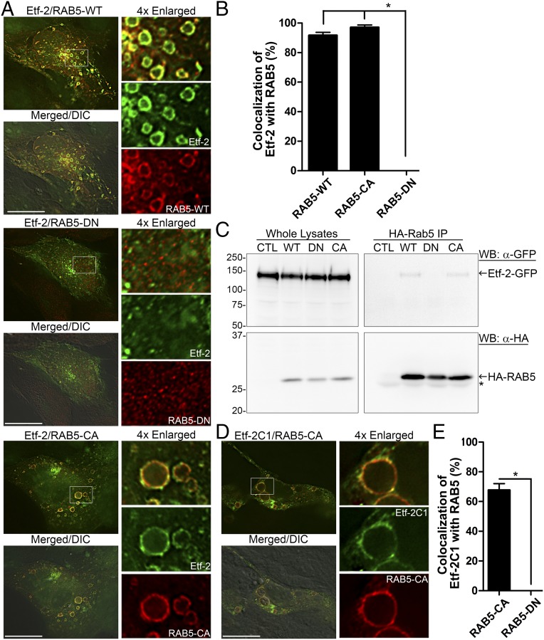
Etf-2 localizes to the early-endosome membrane and interacts with RAB5-GTP. (A) RF/6A cells cotransfected with Etf-2-GFP and HA-RAB5 (WT, DN, or CA). (B) Quantification of 150 vesicles in 20–30 cells cotransfected with Etf-2 and RAB5 (WT, CA, or DN) from three independent experiments. *P < 0.05, ANOVA. (C) HEK-293 cells were cotransfected with plasmids expressing Etf-2-GFP and HA-RAB5 (WT, DN, or CA) or none (CTL), lysed at 2 dpt, immunoprecipitated with mouse anti-HA–linked protein G-Sepharose beads, and analyzed by Western blotting with anti-GFP and anti-HA. The asterisk indicates the IgG light chain. (D) RF/6A cells were cotransfected with Etf-2C1-GFP and HA-RAB5-CA. In A and D cells were subjected to immunofluorescence labeling with mouse anti-HA (red; AF555) at 2 dpt. Merged/DIC, the fluorescence image is merged with the DIC image. Each boxed area is enlarged 4× on the right. (Scale bars: 10 µm.) (E) Quantification of Etf-2C1-GFP colocalization to 100 vesicles or dots of HA-RAB5-CA or HA-RAB5-DN in 30 cells. *P < 0.05, two-tailed t test.

