Fig. 6.
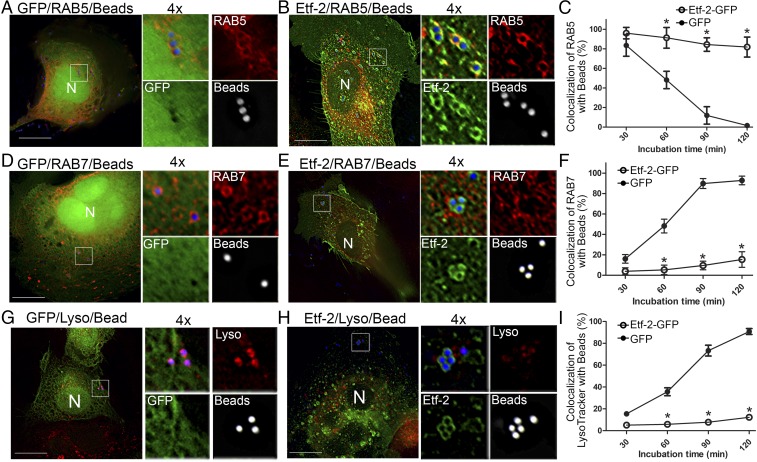
Etf-2 localizes to endosomes containing EtpE-C–coated latex beads and delays the maturation of endosomes in nonphagocytic RF/6A cells. RF/6A cells were cotransfected with GFP (A, D, and G) or Etf-2-GFP (B, E, and H) and HA-RAB5 (A and B) or HA-RAB7 (D and E) for 2 d and then were incubated for 30–120 min with EtpE-C–coated Flash Red latex beads (1 µm, pseudocolored blue for merged images or white for enlarged single-channel panels). At 30–120 min after the addition of latex beads, cells were treated with LysoTracker Red for 10 min before fixation and were immediately observed with a DeltaVision microscope (G and H). In A, B, D, E, G, and H, representative images at 60 min after bead uptake are shown, and each boxed area is enlarged 4× on the right. (Scale bars: 10 μm.) (C, F, and I) Quantification of the localization of RAB5 (C), RAB7 (F), and LysoTracker Red in 100 endosomes that had taken up latex beads (I) in Etf-2-GFP–transfected or GFP-transfected RF/6A cells. Data are presented as the mean ± SD from three independent experiments. *P < 0.05, two-tailed t test.

