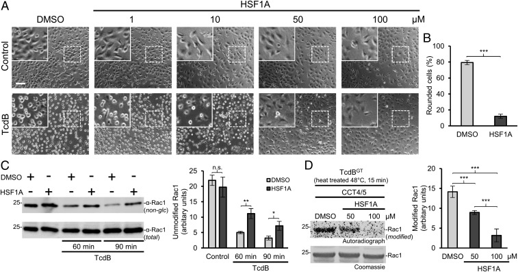
Fig. 2.
HSF1A inhibits TcdB-mediated intoxication of HeLa cells. (A) HeLa cells were pretreated with the indicated concentrations of HSF1A or DMSO as control for 1 h before intoxication with TcdB (5 pM). Pictures were taken after 90 min. (Scale bar: 100 µm.) (Insets) Magnifications of dotted areas. (B) Quantification of HeLa cell intoxication with TcdB (5 pM) after 90 min with pretreatment of DMSO or HSF1A (100 µM). The percentage of rounded cells per picture (>500 cells) is given as mean ± SD (n = 4). Student’s t test was applied for statistical comparison. ***P < 0.001. (C, Left) HeLa cells, pretreated for 1 h with DMSO (control) or HSF1A (100 µM), were incubated with or without TcdB (5 pM) for 60 or 90 min. Cell lysates were analyzed by Western blot with the specific anti-Rac1 antibody (MAB102), which does not recognize glucosylated Rac1. Anti-Rac1 (23A8) antibody served as input control. (C, Right) Quantification of three experiments. Values are average ± SD. Student’s t test was applied for statistical comparison. n.s., not significant (P > 0.05); *P < 0.05; **P < 0.01. (D, Left) CCT4/5-mediated recovery assay of heat-treated (48 °C) TcdBGT with the addition of DMSO or HSF1A (50 or 100 µM). Shown is the [14C]glucosylation of Rac1. (D, Right) Quantification of four experiments. Values are average ± SD. Student’s t test was applied for statistical comparisons. ***P < 0.001.

