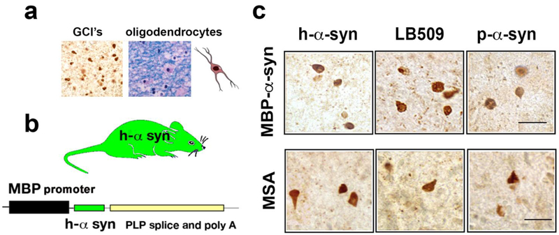
Figure 1.
Comparison of α-synuclein accumulation in oligodendroglial cells in the MBP-α-syn and in MSA. (a) α-synuclein accumulation (left panel) in oligodendroglial cells forming glial cytoplasmic inclusions (GCI’s). Luxol fast blue staining of myelin and oligodendrocytes in MSA brain (right panel). (b) Comparison of α-synuclein inclusions in the MBP model and MSA, images are from the white matter tracts in the striatum immunostained with antibodies against h-α-synuclein, misfolded α-synuclein (LB509 clone) and p-Ser129-α-synuclein. Bar=25 μm. (c) Schematic representation of the MBP transgenic mouse model of MSA driving human α-synuclein.

