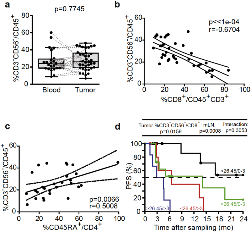
Figure 5. Tumor CD3-CD56- cells inversely correlated with intratumoral cytotoxic T lymphocytes (CTLs) and negatively predicted progression-free survival (PFS).

(a) Flow cytometric analyses of blood and tumor CD3-CD56- cells performed in a paired manner. (b, c) Spearman correlations between CD3-CD56- cells and (b) CD8+ CTL tumor-infiltrated lymphocytes and (c) CD4+CD45RA+ tumor-infiltrated lymphocytes. Each dot represents one patient. Wilcoxon paired signed-rank and Spearman correlation tests: P-values are indicated. (d) Kaplan-Meier PFS curves segregating the cohort of metastatic melanoma patients according to the median value of tumor CD3-CD56- cells retained in the Cox multivariate model in tumor analyses. Likelihood ratio tests from Cox regression modeling are used to assess the prognostic value for the marker with and without accounting for metastatic lymph node (mLN) invasion status (0–3 vs >3).
