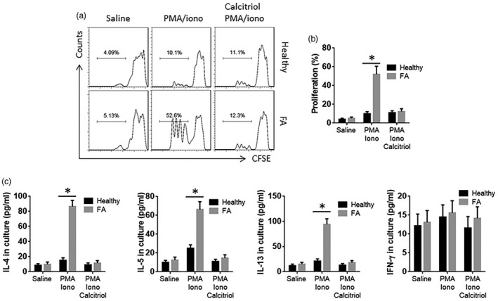
Figure 2.

CD4+ T cells from food allergy (FA) patients show hyperreaction to external stimuli. CD4+ T cells were isolated from peripheral blood mononuclear cells (PBMC) of healthy subjects (n = 6) and FA patients (n = 6). The cells were cultured in the presence of saline, phorbol myristate acetate (PMA) (10 ng/ml)/ionomycin (iono; 100 ng/ml) or PMA/ionomycin/calcitriol (10 nM) for 3 days. (a) The cells were labelled with carboxyfluorescein succinimidyl ester (CFSE) before the culture. The histograms show the proliferation of CD4+ T cells. (b) The bars show the summarized data of panel (a). (c) The bars show the cytokines in the culture supernatant [by enzyme‐linked immunosorbent assay (ELISA)]; *P < 0·01 (t‐test). The samples from individual subjects were analysed separately. Each sample was analysed in duplicate.
