Figure 4.

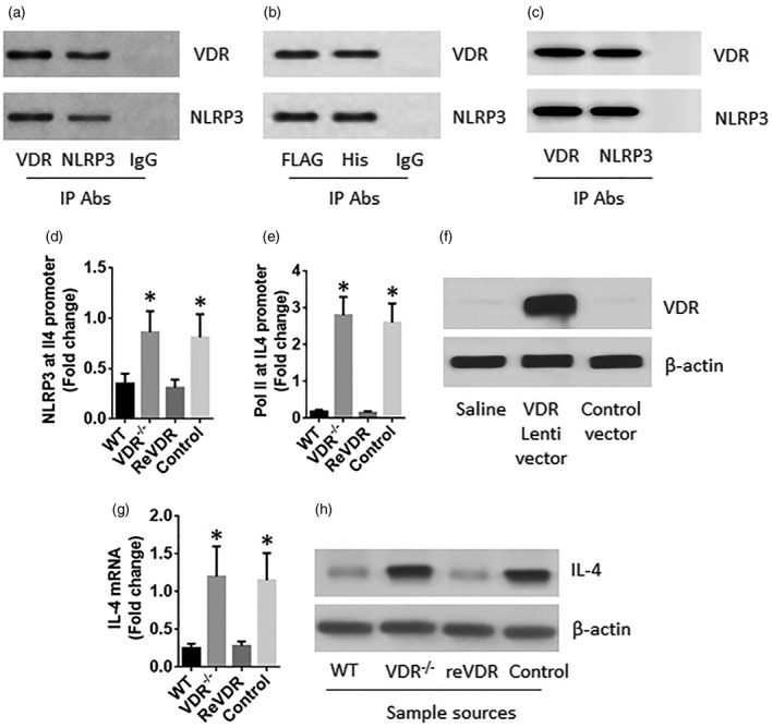
Vitamin D receptor (VDR) restricts interleukin (IL)‐4 expression in CD4+ T cells. (a) A complex of VDR and nucleotide‐binding, oligomerization domain (NOD)‐like receptor family, pyrin domain containing 3 (NLRP3) in wild‐type (WT) CD4+ T cells. (b) A complex of VDR and NLRP3 in human embryonic kidney (HEK)293 cells after transfection of VDR‐FLAG plasmids and NLRP3‐his plasmids. (c) A complex of recombinant VDR and recombinant NLRP3 formed in vitro. (d–h) CD4+ T cells were isolated from the spleen of WT and VDR–/– mice. A portion of the VDR–/–CD4+ T cells were transduced with the VDR gene‐carrying lentivectors or control vectors. (d,e) The nuclear extracts of the cells were analysed by chromatin immunoprecipitation (ChIP). The bars indicate the levels of NLRP3 and RNA polymerase II (Pol II) at the IL4 promoter locus. (f) The results of VDR over expression in CD4+ T cells. (g,h) The expression of IL‐4 in the cells. The data of bars are presented as mean ± standard deviation (s.d.); *P < 0·01 (t‐test), compared with the WT group. The data represent three independent experiments.
