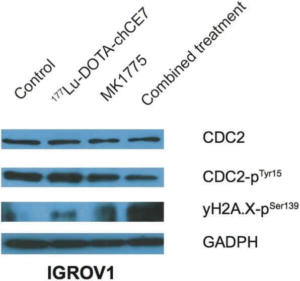
Fig. 3.

Western blot analysis of CDC2, phosphorylated CDC2 and phosphorylation of H2A.X in IGROV1 cell lysates. Cells were treated with either 5 MBq/ml 177Lu-DOTA-chCE7 for 8 h or 300 nM MK1775 for 48 h. For combination, cells were simultaneously incubated with both agents. After incubation (8 h) 177Lu-DOTA-chCE7 was removed and MK1775 further incubated until 48 h were reached. All cell lysates were taken directly post MK1775 treatment. Loading control: GADPH (glyceraldehyde 3-phosphate dehydrogenase)
