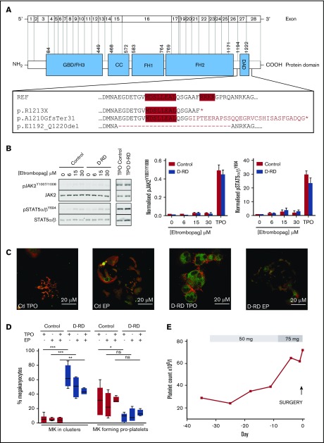
Figure 2.
Detailed evaluation of D-RD cases. (A) Schematic representation of DIAPH1 and DIAPH1 protein divided into functional domains, including the DAD near the C terminus. The expanded box shows the wild-type DAD amino acid sequence; the positions of the regulatory RRKR and MDxLLExL sequence motifs are indicated by red shading. The predicted impact of the variants associated with D-RD is shown compared with the reference sequence. The abnormal C terminus amino acid sequence predicted from the p.A1210GfsTer31 variant is indicated in red type. The position of exon 27 residues that are absent with the p.E1192_Q1220del variant is indicated by the dashed line. *Premature stop codon. (B) Representative immunoblot using monoclonal antibodies recognizing p-STAT5α/βY694, total STAT5α/β, pJAK21007/1008, and total JAK2 of lysates from case A-2 and control platelets stimulated with eltrombopag (0-30 μM) or TPO (100 ng/mL) (left panels). Bar graphs of the ratio of phosphorylated/total densitometry signal of 3 D-RD cases (A-2, B-4, and C-9) combined show that eltrombopag causes markedly reduced STAT5α/β and JAK2 phosphorylation compared with TPO and that the extent of phosphorylation in D-RD platelets is the same as controls (middle and right panels). The data are representative of 3 independent experiments expressed as mean ± standard error of the mean. (C) Representative immunofluorescence confocal microscopy images (case A-2 and control) of differentiated peripheral blood–derived CD34+ MKs at day 12 of culture, visualized using anti-integrin β3 (green; CD61) and phalloidin (red; F-actin) staining. In the presence of TPO, D-RD MKs show abnormal clustering, reduced proplatelet formation, and abnormal distribution of F-actin when compared with controls. Reduced proplatelet formation and cluster formation are partially rescued in culture conditions containing eltrombopag (EP). (D) Corresponding bar graphs of aggregate data from duplicate MK-differentiation experiments from 3 unrelated healthy controls and cases A-2, B-4, and C-9, with each of the 3 D-RD variants cultured with TPO (5 μM), EP (3 μM), or TPO (2.5 μM) + EP (2 μM). Data are expressed as mean and standard error of the mean of the percentage of all cultured MKs that associate in clusters and the percentage that are forming proplatelet extensions, as specified in supplemental Methods. (E) Time course of the hematological response to oral eltrombopag administered to case C-9 before elective hip arthroplasty at day 0. Platelet counts were determined using a Sysmex XN analyzer using the fluorescence end point. ***P < .0001, **P < .001, *P < .05, 1-way analysis of variance. ns, not significant.

