


Figure 3. Schwann cell associated changes within long and short isografts.
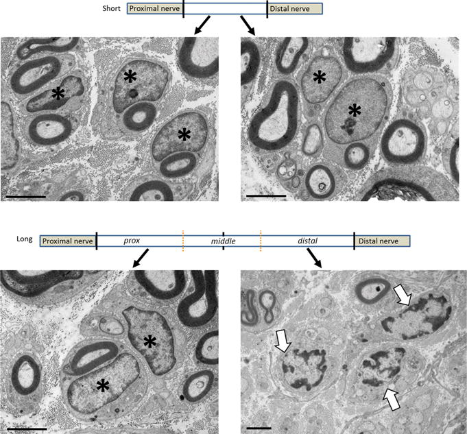
Eight weeks following grafting, electron micrographs of both the proximal and distal portions of the short graft show SCs with normally distributed chromatin around the edges of the nuclei (A). While this chromatin structure is maintained in the proximal aspect of the long graft, the chromatin becomes clumped and scattered within SCs in the distal long graft (B). Scale bar = 200μm. * indicates normal SCs, arrows denote abnormal chromatin