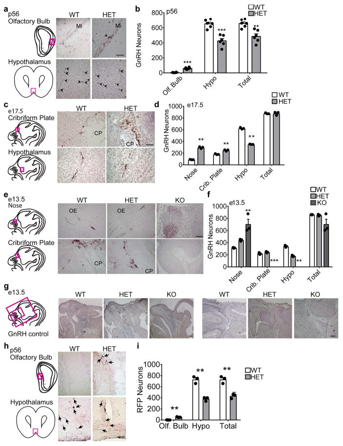
Fig. 2.
Six3 HET and KO mice showed delayed migration of GnRH neurons. Six3 HET and WT mice were processed for IHC staining for GnRH. a, c, and e, Representative images of staining in Six3 WT and HET mice are shown with boxed areas on the drawings of the sagittal mouse head sections to the left of the panels indicating where the (a, c, and e) images were taken. b, GnRH neurons at p56 (Student’s t-test, n=6 (3 females, 3 males)). Counting was conducted throughout the adult brain beginning with the front of the olfactory bulb to bregma −2.80. (olf. bulb, p= 0.00004, t(6)=11.1), (crib. plate, p=0.0001, t(9)=−6.17), (total, p=0.002, t(8)=−4.34). d, GnRH neurons at e17.5 (Student’s t-test, n=3). Counting was conducted throughout the entire embryonic head (nose, p=0.001, t(2)=−22.4; crib. plate, p=0.003, t(4)=−7.40; hypo, p=0.0004, t(2)=31.1; total, p=0.973, t(2)=−0.03). f, GnRH neurons at e13.5 (Student’s t-test, n=3; nose, p=0.002, t(3)=−12.2; crib. plate, p=0.116 t(4)=−2.06; hypo, p=0.0002, t(4)=13.6; total, p=0.488, t(2)=0.821). g, Controls for GnRH staining specificity, images are from e13.5 mice processes for anti-GnRH IHC. h and i, lineage tracing, IHC for RFP in Six3HET:RosaRFP:GnRHcre adult mice. RFP marks all GnRH neurons for the duration of the life of the neuron regardless of GnRH expression, to determine presence of GnRH neurons lacking GnRH expression. i, Number of RFP-labelled GnRH neurons in Six3HET:RosaRFP:GnRHcre adult mice. (Student’s t-test, n=3; olf. bulb, p=0.004, t(2)=−14.0; hypo, p=0.003, t(3)=8.41; total, p=0.004, t(3)=6.87). Arrows indicate RFP neurons. Mi, mitral layer; CP, cribriform plate; OE, olfactory epithelium. **p<0.005, ***p<0.001. Scale bars, 100 μm (a, c, e, h), 2 mm (g left), 200 μm (g right).

