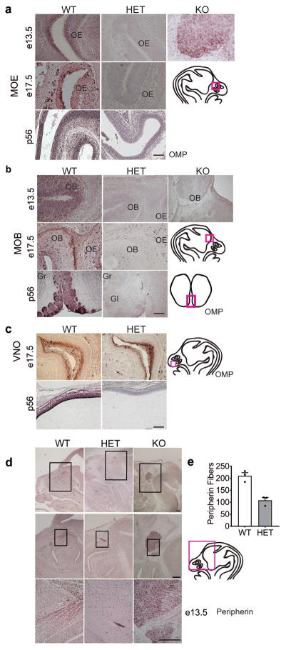
Fig. 7.
Six3 HET mice lack olfactory neurons. a, b, and c, Olfactory Marker Protein (OMP) IHC to mark all mature olfactory sensory neurons (OSNs) of Six3 WT, HET and KO mice at e13.5, 17.5, and p56 in (a) the MOE, (b) the MOB, and (c) the VNO, n=3. Boxes on drawings of the brain to the right of representative IHC images indicate where the pictures were taken. d, IHC for Peripherin-positive axons in the olfactory system at e13.5 in Six3 WT, HET, and KO mice, n=3. e, quantification of peripherin fibers in Six3 WT and HET embryos (Student’s t-test, p=0.004, t(4)=5.82, n=3). Boxes on drawings of adult brain (a) or embryo head (b, c) indicate where the representative images were taken. OB, olfactory bulb; OE, olfactory epithelium; Gr, granular layer; Gl, glomerular layer. Scale bars, 100 μm (a, b, c), panel d, 2 mm, 10 μm, 100 μm.

