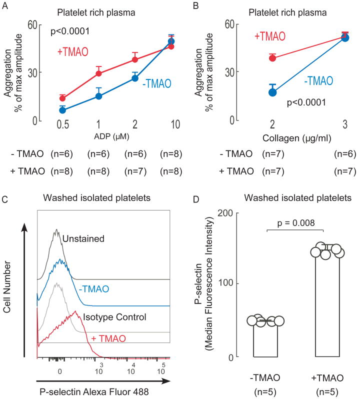
Figure 1. Brief exposure of platelets to TMAO enhances platelet responsiveness.
(A–B) Platelet rich plasma (PRP) was recovered from C57BL/6J female mice (maintained on chow diet) and then incubated in the presence of TMAO (100 μM) versus vehicle (normal saline, “-TMAO”) control for 15 min as described under Methods. Platelet aggregometry response following stimulation with the indicated concentrations of either ADP (A) or collagen (B) were then examined. Data represent the % of maximum aggregometry amplitude (mean ± SD) from the indicated number of distinct biological replicates (mice). (C–D) Platelets were isolated from C57BL/6J female mice maintained on chow diet, washed, and then incubated with either TMAO (100μM) or vehicle (normal saline) as described under Methods. At time of exposure to ADP (1 μM), either anti P-selectin or isotype control antibody were concurrently added, and then following 10min, cells were fixed and surface expression of P-selectin was quantified by flow cytometry, as described under Methods. Illustrative flow cytometry tracings (C) and quantitative P-selectin surface expression (median fluorescence intensity, ± SD) from the indicated number of mice (D) are shown. P values shown were calculated using Kruskal-Wallis test( A,B) and Wilson rank-sum test was used for two-group comparison (D).

