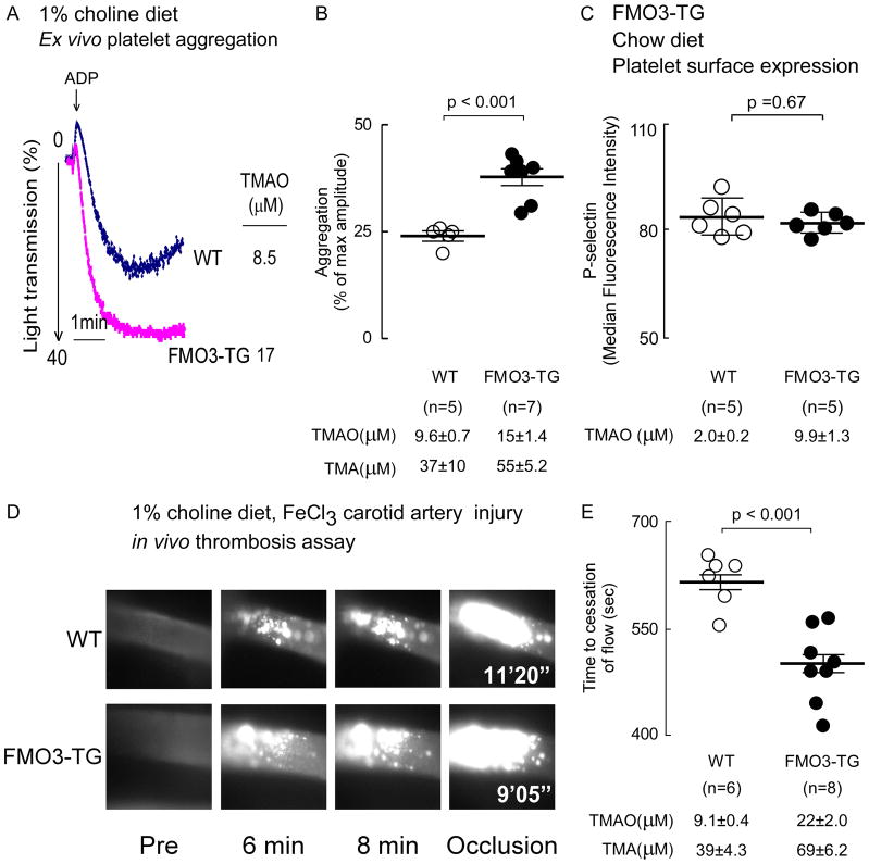
Figure 6. Over expression of human fmo3 transgene in mice leads to increased platelet responsiveness and enhanced thrombosis potential in vivo.
(A, B) Platelets rich plasma was isolated from human liver specific FMO3-TG mice and their WT littermates maintained on a choline (1%) supplemented diet. Plasma TMA and TMAO levels were quantified by stable isotope dilution LC/MS/MS analysis, and platelet aggregometry was monitored following stimulation with sub-maximal (1μM final) ADP, as described under Methods. (A) Representative aggregation tracings in response to ADP from a mouse within each group, along with plasma TMAO level in the mouse. (B) Cumulative data from mice in each group. Data represent the % of max aggregometry amplitude in response to 1μM ADP, along with mean (±SEM) plasma TMA and TMAO levels, for the indicated mouse groups. (C) Washed isolated platelets were recovered from hepatocyte-specific FMO3-TG mice and their WT littermates maintained on a chow (non-supplemented with choline) diet, and then surface expression of P-selectin in response to submaximal ADP (1 μM) was determined by flow cytometry, as described in Methods. P-selectin surface expression (median fluorescence intensity, mean ± SD) from the indicated number of mice are shown. (D, E) Vital microscopy images and quantified time to cessation of blood flow in carotid artery following FeCl3-induced carotid artery injury in the indicated groups of mice. (E) Plasma TMA and TMAO levels are also shown. For all panels, the middle line represents mean and whiskers represent SEM for the indicated numbers of mice. The Wilcoxon rank-sum test was used for two-group comparison.

