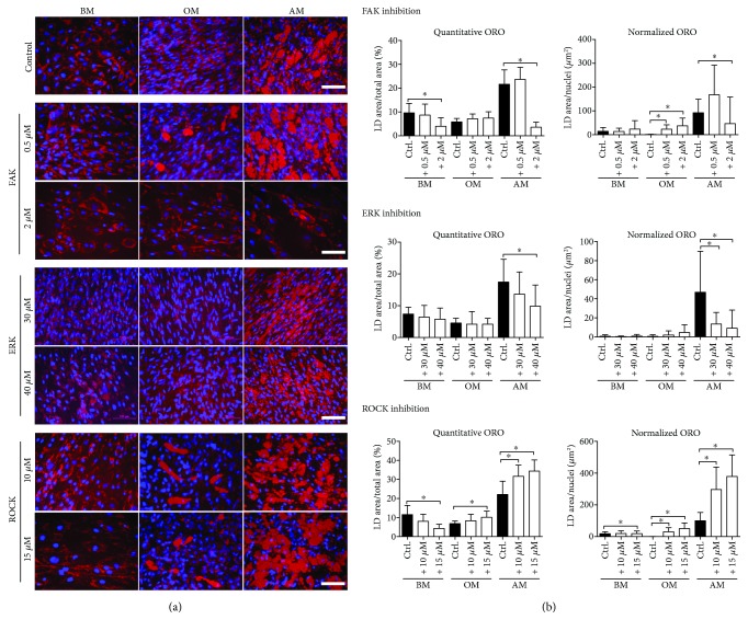
Figure 5.
ORO staining of hASCs and quantification of lipid accumulation from ORO-stained fluorescence images. (a) Representative ORO- and DAPI-stained fluorescence images of FAK, ERK, and ROCK inhibitor-treated hASCs at 21 d. Human ASCs were stained with ORO for intracellular lipid accumulation followed by nuclei staining with DAPI. Fluorescence images were taken with Alexa546 for ORO (red) and DAPI (blue) filters. Scale bars 100 μm. (b) ORO-stained samples of FAK, ERK, and ROCK inhibitor-treated hASCs were imaged with fluorescence microscope using Alexa546 and DAPI filters and analyzed with a custom analysis pipeline designed for CellProfiler. Quantitative ORO graph presents the area of all stained LDs as percentages of the total image area. Normalized ORO graph describes LD formation on the single cell level: the area of LD clusters over 10 μm in diameter is normalized with the corresponding nuclei count. Significance level 5%, designated with an asterisk (∗). FAK, ROCK: N = 13–16 (images from 2 donors), ERK: N = 19–21 (images from 3 donors). BM: basic medium; OM: osteogenic medium; AM: adipogenic medium; ORO: Oil Red O; LD: lipid droplet.

