Fig. 2.

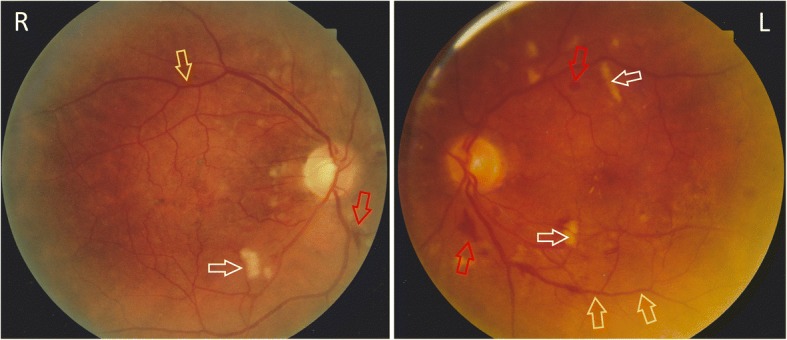
Bilateral fundus colour photographs demonstrating cotton wool spots (white arrows), venous beading (yellow arrows) and haemorrhaging (red arrows), consistent with hypertensive retinopathy

Bilateral fundus colour photographs demonstrating cotton wool spots (white arrows), venous beading (yellow arrows) and haemorrhaging (red arrows), consistent with hypertensive retinopathy