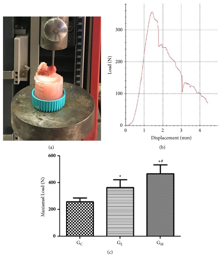
Figure 5.
(a) Illustrating how the specimen was fixed in dental cement and the force was parallel to the long axis of the proximal femur. (b) Representative load-deformation curve obtained by the biomechanical test. (c) Statistical analyses of the biomechanical test data. ∗p<0.05 vs. the blank group; #p<0.05 vs. GL.

