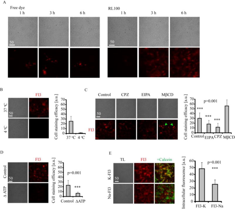
Figure 4. Mechanism of cell entry for FI3 nanoparticles.

A: Staining kinetics for FI3 (0.1 μM free dye or 10 μg/ml RL100 nanoparticles) in live rat primary neural cells. Cells were incubated with FI3 for indicated time intervals, washed and immediately imaged. B-D: Staining of primary neural cells with FI3 nanoparticles (10 μg/ml, 3 h). B: Effect of temperature on staining efficiency. C: Effects of various endocytosis inhibitors on cell staining with FI3. Cells were pre-treated with inhibitors (50 μM EIPA, 10 μg/ml CPZ and 5 mg/ml MβCD) for 30 min, followed by staining procedure. MβCD-treated cells displayed round morphology, indicated with green arrows. N= 17. D: Effect of ATP depletion on cell staining efficiency. For ATP depletion, cells were pre-incubated in no-glucose medium and treated with oligomycin (10 μM), followed by staining procedure. N=20. E: Cell staining efficacy for K-FI3 and Na-FI3. Cells were incubated with nanoparticles (10 μg/ml, 16 h) washed, counter-stained with Calcein Green and imaged. Scale bar is in μm.
