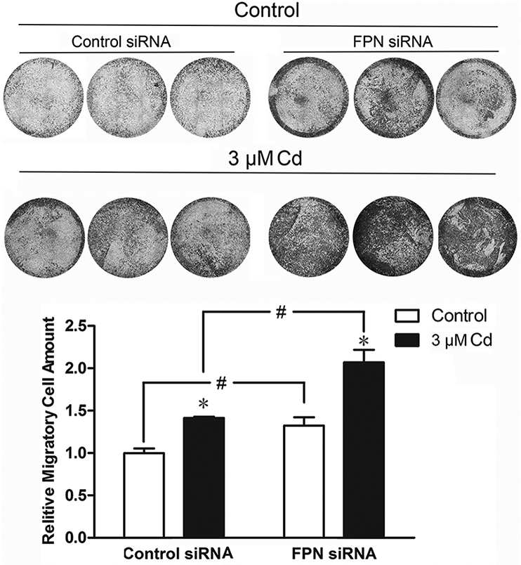
Fig. 6.

Effect of suppression of FPN expression on the migration of cells treated with Cd. The cells were treated with 3 μM Cd for 8 weeks. Control cells were cultured in DMEM alone. Cells were transiently transfected with either control or FPN siRNA for 2 days and seeded in Transwell inserts. After 48 h, migrated cells were imaged after staining with crystal violet. Relative migration ability was quantified by measuring the absorbance of the de-staining solution. Results are expressed as mean±SE (n=3). *Significantly different from the respective control.#Significantly different from the cells treated with control siRNA. (p < 0.05).
