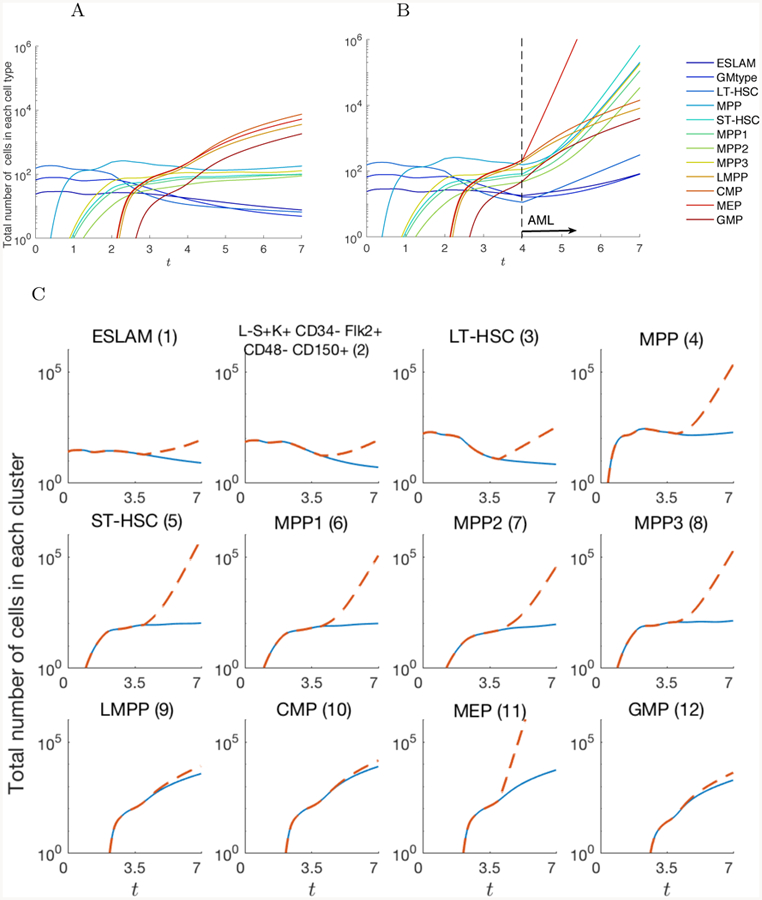
Figure 7.

Total number of cells in each node up to t = 7 in A) normal condition and B) AML pathogenesis. The AML simulation is started at t = 4. Compared to the normal case, cells in MEP, LT-HSC, and ST-HSC increase as well as other cell types. Figure C) compares the number of cells between the normal and AML case for each cell type individually.
