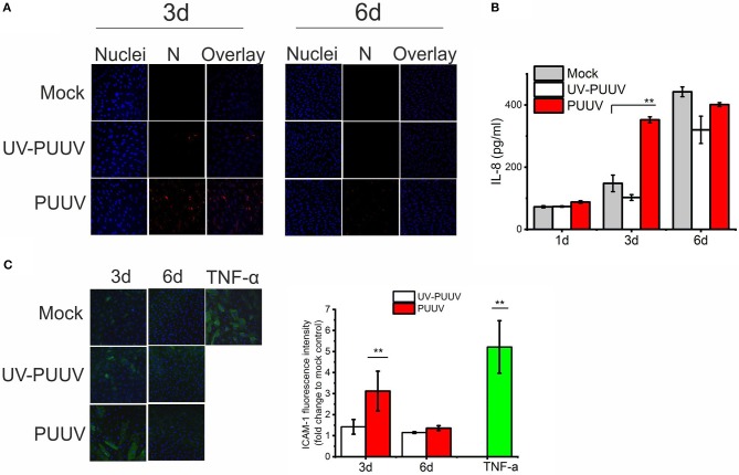
Figure 4.
PUUV infection causes pro-inflammatory responses in BECs. (A) BECs were either mock-infected or infected with live or UV-inactivated PUUV (UV-PUUV) and assessed for viral nucleocapsid protein expression at 3 and 6 days post infection by immunofluorescence (red). The nuclei of BECs were visualized with Hoechst 33420 (blue). (B) IL-8 was measured from the respective supernatants of mock, UV-PUUV or PUUV-infected BECs by ELISA. (C) ICAM-1 expression in TNF-α treated or mock-, UV-PUUV or PUUV-infected BECs was visualized by immunofluorescence (green) and shown as an overlay with Hoechst 33258 staining (blue). Fluorescence intensity of ICAM-1 expression on BECs was quantified and reported as fold change to mock-infected cells. Differences between groups were assessed by one-way ANOVA with Dunnett's multiple comparisons test and statistically significant differences indicated as **p < 0.01. n = 2 in all panels. Results shown are representatives of three independent experiments.

