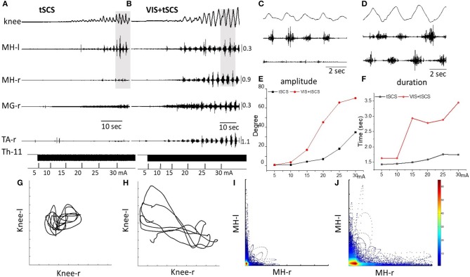
Figure 2.
Angular excursions of the right knee joint and EMG activity in the medial hamstring (MH) in right and left legs, right medial gastrocnemius (RMG), and right tibialis anterior (RTA) during gradual (by 5 mA) increasing intensity of spinal stimulation alone (A) and in the presence of visual imagery of stepping (VIS) (B) in Subject D.G. Knee excursion and EMG bursts marked by a gray background (A,B) are displayed with an extended time scale in (C,D), correspondingly. Plots of amplitude displacements (E) and cycle period (F) of knee joint during tSCS alone and VIS+tSCS. Kinematics coordination based on knee (left) and knee (right) movements during tSCS (G) and VIS+tSCS (H). Pattern of reciprocity for EMG activity of the HM (left) and HM (right) during tSCS (I) and VIS+tSCS (J). EMG calibration: mV.

