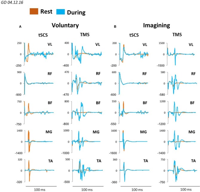
Figure 7.
Averaged sEMP during tSCS delivered at L1 and MEPs during TMS, recorded in mm. vastus lateralis (VL), rectus femoris (RF), medial hamsting (MH), medial gastrocnemius (MG) and tibialis anterior (TA) in one participant at rest (orange) and during voluntary stepping (blue) (A) as well as during visual imagery stepping (blue) (B).

