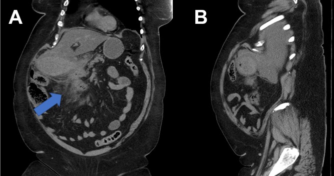
Figure 2.

(A and B) Coronal and axial films of CT abdomen/pelvis demonstrating the thickened gallbladder wall with high density and induration of the surrounding fat (thick arrow) extending into the porta hepatis and adjacent to the second part of duodenum.
