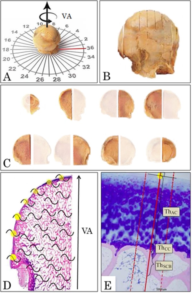
Figure 1.

Design-based stereological methods using systematic uniform random sampling and vertical sections were used to study the undecalcified human femoral heads. (A) The vertical arrow represents the vertical axis (VA) of the femoral head and the rotated arrow represents the random rotation of the femoral head around the VA. The red line indicates the location of the medial part of the femoral head. (B) The femoral head was cut in 7 mm thick slices in a random orientation and parallel to the VA to get vertical uniform random sections. (C) The 7 mm thick slices were halved and alternating left and right half slices were sampled randomly resulting in a sampling fraction of one-half. (D) From each femoral head, all the sampled 7 mm thick halved parallel slices were embedded in methylmethacrylate, and 7 µm thick histological sections were cut from each slice and stained with May-Grünwald toluidine blue. In all histological sections, the joint surface was sampled systematically uniform random as every intersection (yellow dots) between the superimposed cycloid grids (black curved lines) and the joint surface. (E) For each random sampling point, the Osteoarthritis Research Society International (OARSI) grade was determined, and a line was drawn perpendicular to the joint surface. Along the line drawn perpendicular to the joint surface, the ThAC, ThCC and ThSCB were measured. To increase the precision of the ThSCB, two lines separated by 600 µm were drawn parallel to and on each side of the line perpendicular to the joint surface. The subchondral bone (SCB) plate thickness was also measured along these additional two lines, and the average was used as the estimate of the SCB plate thickness at that specific sampling point. AC, articular cartilage; CC, calcified cartilage.
