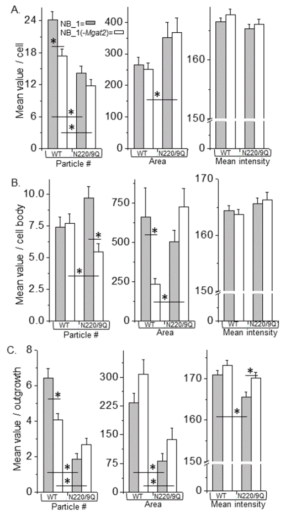
Figure 4:

Comparison of neuronal membrane distribution of Kv3.1b with predominant expression of hybrid or complex types of N-glycans. Fluorescent particles from EGFP tagged glycosylated (WT) and unglycosylated (DM) Kv3.1b proteins in NB_1 and NB_1 (-Mgat2) cells were analyzed in or near plasma membrane of cell (A), cell body (B), and outgrowth (C). Number (left panels), area (middle panels) and mean intensity (right panels) of particles were compared between the glycosylated Kv3.1b proteins, as well as the unglycosylated proteins, in each cell line. The glycosylated and unglycosylated Kv3.1b proteins expressed in the same cell line were also compared. Experiments were performed on at least 3 different days and at least 31 cells were examined in each group. Asterisks indicate significant differences in mean values at a probability of P<0.05 using unpaired student T-test.
