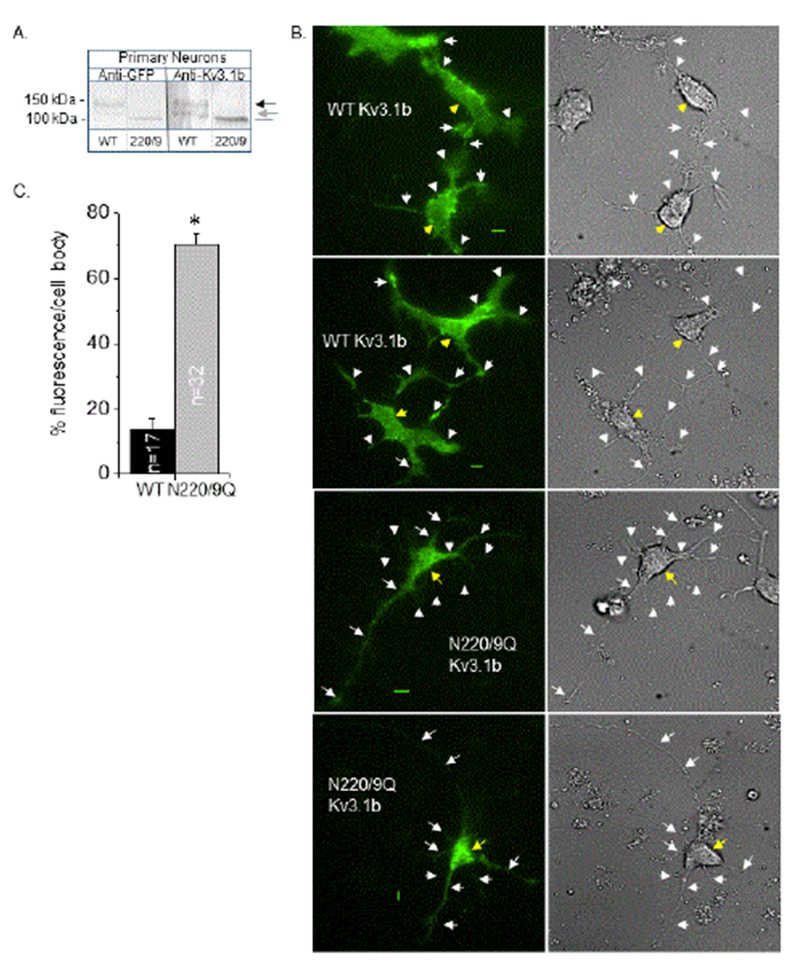
Figure 5:

Characterization of ectopic expression of glycosylated and unglycosylated forms of the Kv3.1b protein in adult primary neurons. Western blots of EGFP tagged Kv3.1b in whole cell lysates from adult primary neurons transfected with glycosylated (WT) and unglycosylated (220/9) forms of the Kv3.1b protein (A). Arrows denote glycosylated forms of the Kv3.1b protein, and gray line represents unglycosylated Kv3.1b protein. Representative TIRF (left panels) and DIC (right panels) images acquired in similar planes are shown for neurons expressing glycosylated (top two panels) and unglycosylated (bottom two panels) forms of the Kv3.1b protein (B). White and yellow arrows point to outgrowths and cell body, respectively. Representative scale bar (5 μm) was identical for image pairs. Percent of fluorescence intensity in cell body was determined to quantify differences between the level of glycosylated and unglycosylated Kv3.1b in cell body (C). Asterisk denotes significant differences in mean values at a probability of P<0.000001 using student t-test. Experiments were performed on at least 3 different days and at least 32 cells were examined in each group.
