
Figure 3. Testing area.
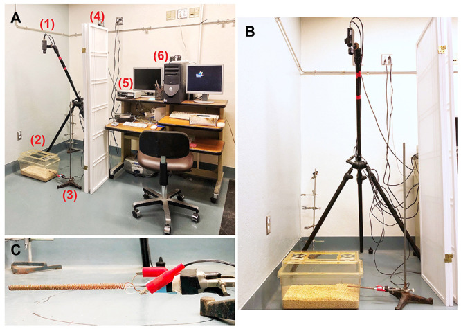
A. Equipment set-up: (1) tripod and camera; (2) cage; (3) ring stand; (4) partition; (5) shock generator; (6) computer; B. Cage positioned under camera, with shock-probe connected to shock generator, clamped to ring stand, and inserted into test cage. C. Close-up of probe clamped to a ring stand, with alligator clips attached.