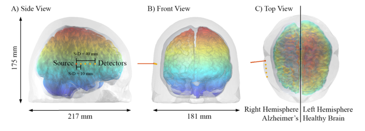
Fig. 4.
The pulsed laser source impinging the human head from its right hemisphere. The detector positions are shown in orange and their distance is with respect each other. In this figure, the right lobe of the brain is from the AD model Stage 6 while the left is the original Colin27 model [31]. To help the visualization of the modeled disease progression, we label the z-coordinate with jet color bar.

