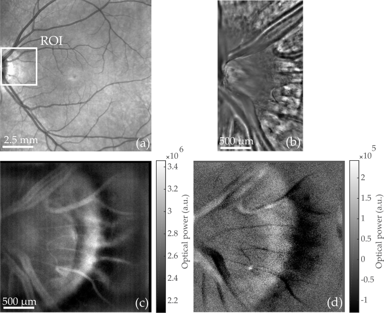
Fig. 3.
Optic Nerve Head (ONH) region imaged with commercialized instruments and Laser Doppler holography. (a) Scanning laser ophthalmoscope (Spectralis, Heidelberg). (b) Adaptive optics flood illumination (rtx1, Imagine Eyes). (c) Power Doppler images M0+ calculated from holograms recorded at fS = 39 kHz; S(ω) is integrated over [ f1, f2] = 4–19.5 kHz. Multiple power Doppler images M0+ are averaged over a total time of 80 ms (see Visualization 1 (101.6MB, avi) for blood flow movie). The peri-papillary crescent is visible on the edge of the ONH. (d) Asymmetry of the DPSD M0− (averaged over the same period of time) illustrating the resultant flow direction with respect to the optical axis.

