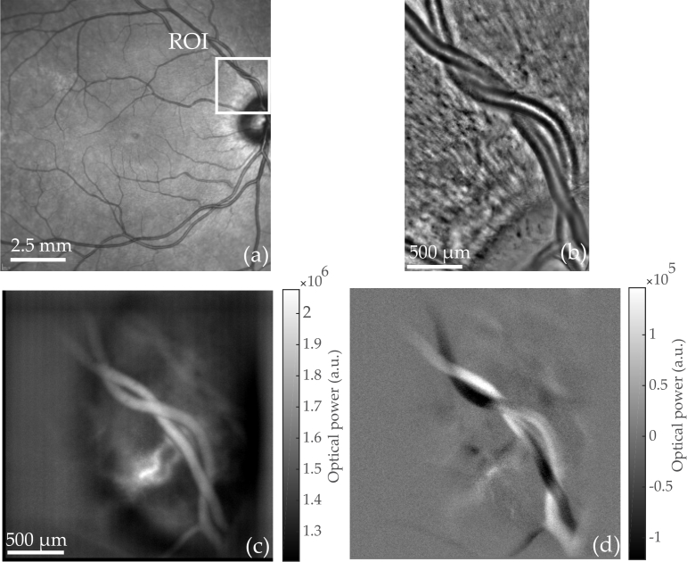
Fig. 4.
An artery intertwined with a vein is imaged with commercialized instruments and Laser Doppler holography.(a) Scanning laser ophthalmoscope (Spectralis, Heidelberg). (b) Adaptive optics flood illumination (rtx1, Imagine Eyes). (c) Power Doppler images M0+ calculated from holograms recorded at fS = 39 kHz; S(ω) is integrated over [f1, f2] = 7–19.5 kHz. Multiple power Doppler images M0+ are averaged over a total time of 0.66 s (see Visualization 2 (96.9MB, avi) for blood flow movie). (d) Asymmetry of the DPSD M0− (averaged over the same period of time) illustrating the resultant flow direction with respect to the optical axis.

