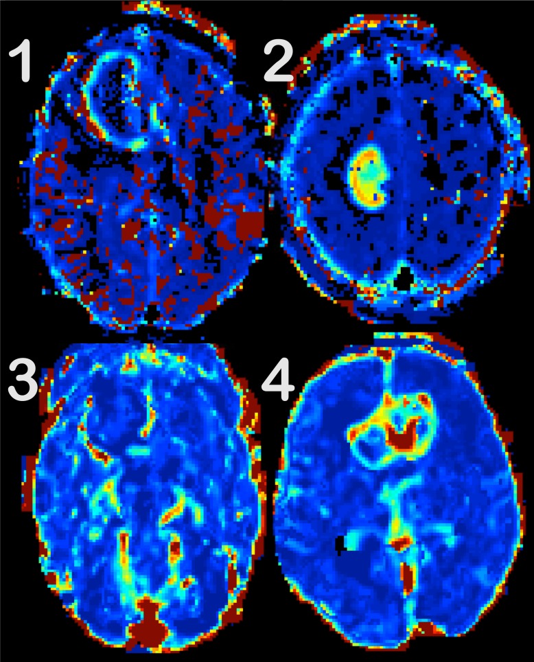
Fig 1. Examples of the 4-step-scale, used for quality assessment.
Tumor-to background discrimination is evaluated for Ve maps. (1) is not interpretable, as spurious voxels (in red and black) partly obscure the tumor. In (2), the tumor is discernible, but still affected by artifacts both in and around the lesion. The image quality is good in (3), but the tumor is difficult to spot due to the surrounding structures and artifacts. The tumor-to-background discrimination is excellent in (4).

