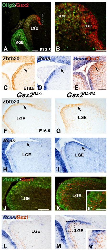
Fig. 7. Candidate glial progenitor genes, Zbtb20 and Bcan are increased in VZ progenitors of Gsx2 mutants.

Immunostaining for 01ig2 and Gsx2 reveals opposing patterns of expression in the E13 LGE (A-B). Dashed box in A depicts the high magnification view of the LGE in B. Gsx2 positive cells in the dorsal LGE region (dLGE) are largely Olig2 negative, whereas double positive cells are throughout the ventral LGE (vLGE). Immunostaining for Zbtb20 (C) and in situ staining for Bcan (D) show weak expression in the dLGE at E18.5 (arrows in C-D). Double labeling with in situ for Bean and IHC for Gsx2 show the dLGE is high for Gsx2 and weak for Bcan (arrow in E). Gsx2 mutants display increased Zbtb20 (compare arrows in G to F) and Bcan (compare arrows in I to H) expression in the dLGE region at E16.5. Immunostaining for Zbtb20 and Gsx1 show the increased Zbtb20 is distinct from the expanded Gsx1 positive cells in VZ progenitors of Gsx2 mutants (compare K to J, see also inset K’). Double labeling with in situ for Bcan and IHC for Gsx1 (L) also shows little overlap in the Gsx2 mutant LGE progenitors (M and M’). Scale bar: A = 200μΜ, B, K% Μ’ = 50μΜ, E 100μΜ for C-E, I = 100μΜ for F- I,L-M, J = 100μΜ for J-K
