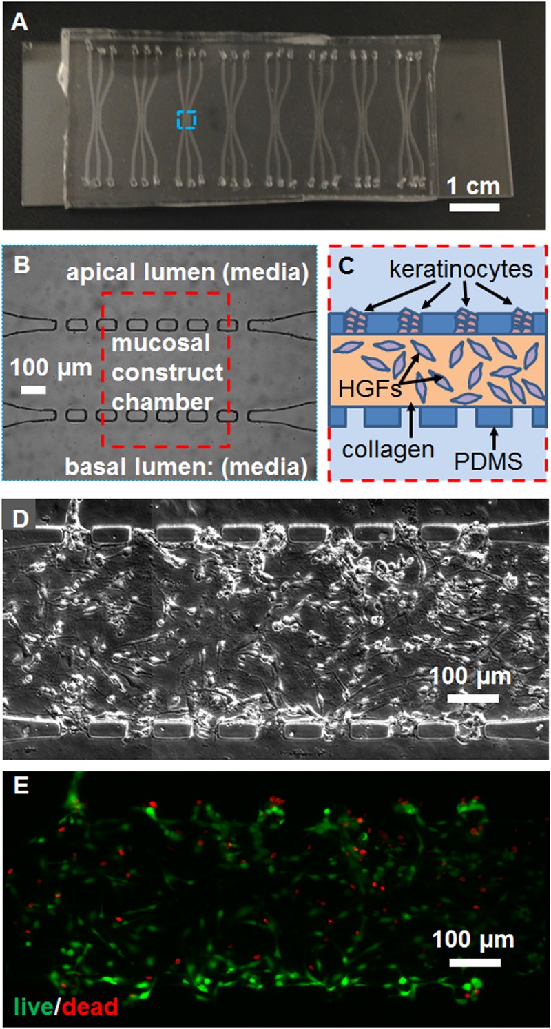
FIG. 1.
Configuration of the microfluidic device and tissue construct. (a) A photograph of the chip containing eight 3-channel devices. (b) A phase contrast micrograph from the blue dashed box in (a), and (c) schematic of the construct culture chamber from the red dashed box in (b), showing channel and pore microstructure and cell placement. Mosaicked (d) phase contrast and (e) epifluorescence micrographs of the live-dead stained co-culture, showing the morphology of cells, viable cells in green, and dead cell nuclei in red. The scale bar is indicated.

