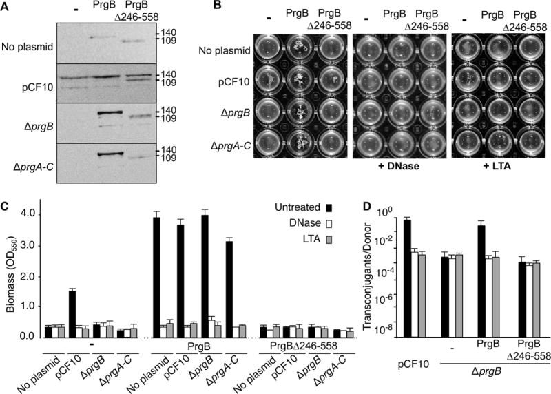
Figure 7.

Contributions of the PrgB adhesin domain to aggregation, biofilm formation, and conjugation. In all panels, strains analysed were OG1RF alone (denoted ‘no plasmid’) or with pCF10 or pCF10ΔprgB (ΔprgB); these strains also were engineered to produce native PrgB or PrgBΔ246-558 from the constitutive P23 promoter. A: Steady-state levels of Prg proteins in strains induced for 1 h with cCF10 pheromone (10 ng ml−1). Immunoblots were developed with anti-PrgB antibodies. Protein sizes (in kilodaltons, kDa) are listed at the right. Protein extracts were loaded on a per-cell equivalent basis. B: Photographs of strains grown in microtiter plates showing the effects of DNase and LTA on PrgB-mediated clumping. C: Biofilm formation on polystyrene microtiterplates. The biofilm biomass was assayed as a function of crystal violet stain retained. DNase or LTA treatment was initiated at the onset of cCF10 induction and biofilm formation and continued for the full incubation time of 24 h. Results are expressed as OD550 values and values represent the average of at least three independent experiments; the error bars represent standard deviations. D: Effect of the PrgB adhesin domain on plasmid transfer. Transfer frequencies are presented as the number of transconjugants per donor cell. Experiments were repeated at least three times in duplicate, and results from a representative experiment are shown.
