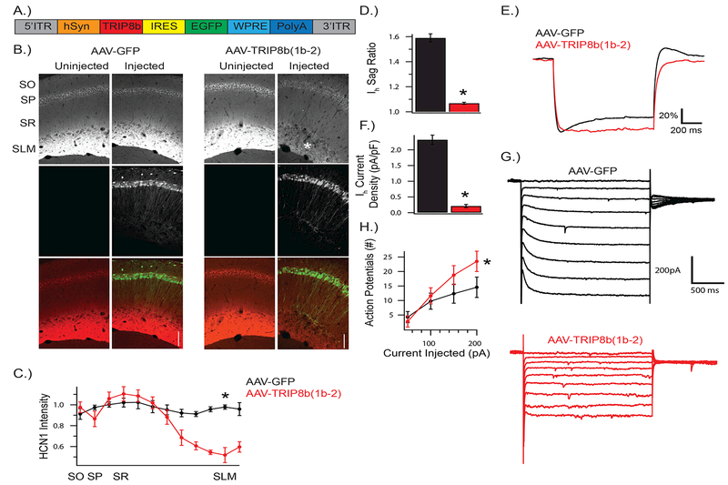
Figure 2: AAV-(1b-2) reduces HCN channels in the CA1.
A) Schematic of AAV-(1b-2). TRIP8b(1b-2) is driven by a human synapsin (hSyn) promoter with IRES-GFP to track infected cells. AAV-GFP expresses GFP through hSyn and lacks TRIP8b(1b-2). B, C) HCN1 is reduced in CA1 cells, especially at the distal dendrites (t4 = 7.712, p < 0.01; nsubjects = 3,3). Asterisk highlights SLM, where HCN1 is reduced after AAV-(1b-2) injection. D,E) Membrane response to a 1s long −200pA current injection from a resting potential of −70mV. Voltage shown is scaled to ∆Vmax to facilitate comparison of sag ratio amongst conditions. AAV-(1b-2) injected cells show reduced sag ratios compared to AAV-GFP (t14 = 14.9, p < 0.001, ncells = 8, 8). and F,G) Ih current density (t13 = −12.0, p < 0.001, ncells = 8, 7), and H) increased excitability (Repeated Measures ANOVA; genotype × stimulus interaction F3,42 = 8.44, p < 0.001, ncells = 8, 7). SO: stratum oriens, SP: stratum pyramidale, SR: stratum radiatum, SLM: stratum lacunosum moleculare.

