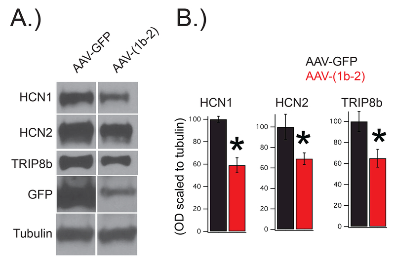
Figure 3: AAV-(1b-2) injection leads to a reduction in HCN1, HCN2, and TRIP8b protein.
Wild type mice were bilaterally injected with either AAV-(1b-2) or AAV-GFP. Four weeks later their hippocampi were sub-dissected for western blot. A.) Representative images. B.) Quantification of the results reveal reduction in HCN1 (AAV-GFP:100±2.7%, AAV-(1b-2: 59±6.7%,nsubjects = 9,12, t19=4.93, p<0.01), HCN2 (AAV-GFP:100±12.3%, AAV-(1b-2: 69±5.7%,nsubjects = 8,11, t17=2.45, p<0.05), and total TRIP8b (AAV-GFP:100±9.5%, AAV-(1b-2: 65±8.5%,nsubjects = 10,12, t20=2.76, p<0.05) in the AAV-(1b-2) injected mice compared to the AAV-GFP controls.

