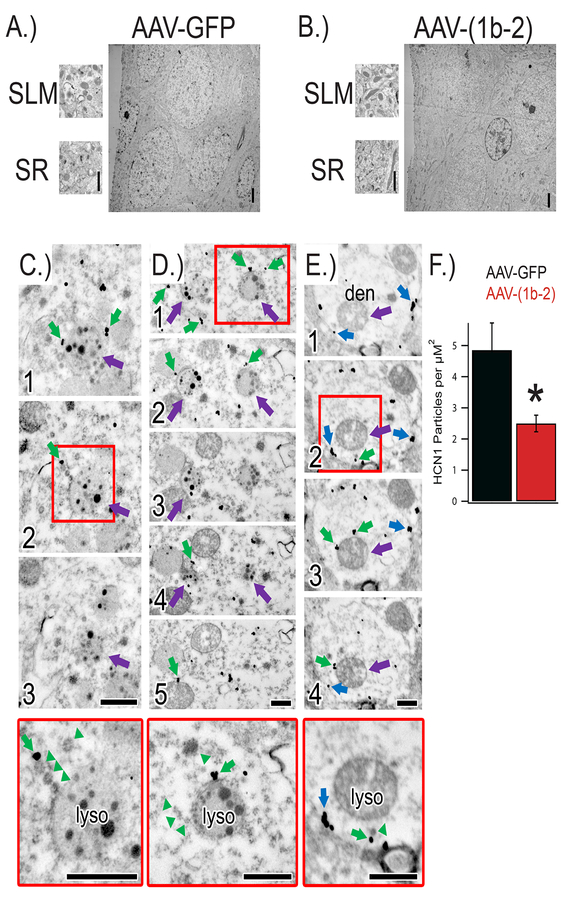
Figure 4: AAV-(1b-2) re-localizes HCN channels to lysosomes for degradation.
A,B) Electron micrographs of hippocampi injected with either AAV-GFP or AAV-(1b-2). C) Electron micrographs of serial sections (1, 2, 3) through a lysosome near the soma (purple arrow) in close proximity to endosomes immunoreactive for HCN1 (immunogold particles; green arrows) in the AAV-(1b-2) injected hippocampi. Red box denotes approximate region shown at higher magnification in bottom panel. Note the ordered manner in which the endosomes are lined up near the lysosome (green arrowheads). D) Electron micrographs of serial sections (1, 2, 3, 4, 5) through two lysosomes near the soma (purple arrows) with endosomes immunoreactive for HCN1 (green arrows) nearby. E) Electron micrographs of serial sections (1, 2, 3, 4) through a spiny dendrite in the stratum lacunosum-moleculare. Despite a dramatic HCN1 decrease in the distal dendrites and the majority of HCN1 immunoreactivity localized to endosomes (green arrows) near lysosomes (purple arrows), occasional cell membrane-bound HCN1 was noted (blue arrows). Higher magnification images reveal orderly queuing near the lysosome (green arrowheads). Scale bars = 500nm. F.) Quantification of HCN1 particle density in the SLM. AAV-GFP 4.85±0.87 particles/μm2 dendrite, AAV-(1b-2) 2.49±0.26, *p<0.05 two tailed T test, nsubjects = 2, ndendrites = 22,23.

