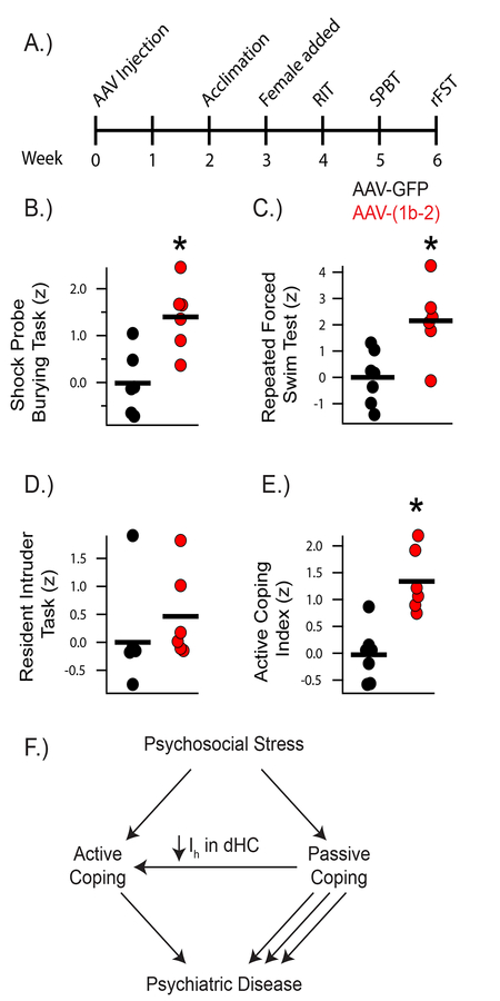
Figure 5: AAV-(1b-2) injection in the CA1 leads to active coping.
A) Timeline of viral injections and Active Coping Index (ACI) behavioral assays. B) AAV-(1b-2) mice showed increased active coping in the Shock Probe Burying Test (AAV-GFP: −0.01±0.27, AAV-(1b-2): 1.39±0.29, t10 = 3.510, p < 0.01, nsubjects = 6, 6). C) AAV-(1b-2) mice showed decreased average immobility time across the three repeated Forced Swim Test trials (AAV-GFP: 172.38±10.48s, AAV-(1b-2): 112.61±16.00s t11 = 3.212, p < 0.01, nsubjects = 7, 6). D) AAV-(1b-2) mice did not demonstrate statistically different z-scores from AAV-GFP mice in the Resident Intruder Test (AAV-GFP: 0±0.40, AAV-(1b-2): 0.46±0.32, t10 = 0.8968, p > 0.05). E) AAV-(1b-2) mice show enhanced active coping compared to AAV-GFP mice on the ACI (t11 = 4.586, p < 0.001, nsubjects = 7, 6). F) Working model of how HCN Channel loss in the hippocampus leads to increased active coping. When an individual encounters acute psychosocial stress, they cope passively or actively. Those with Active Coping styles tend to be more resistant to developing most neuropsychiatric disorders and tend to do better with treatment than those who employ Passive Coping styles. Reduction of HCN channels and HCN channel current (Ih) specifically in the CA1 facilitates Active Coping strategies.

