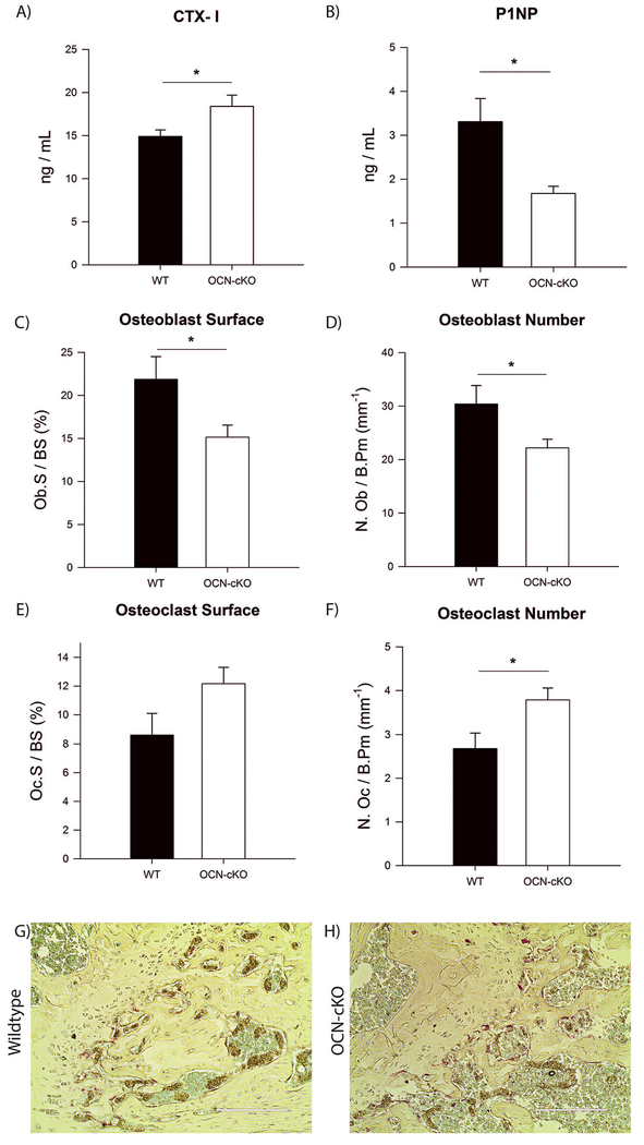
Fig. 2.
Bone formation is decreased and resorption is increased due to a reduced number of osteoblasts and an increased number of osteoclasts, respectively. (A) Serum from WT and OCN-cKO mice was assayed using RatLaps (C-terminal telopeptides of type I collagen, CTX-I) EIA. (B) Serum from WT and OCN-cKO mice was assayed using P1NP EIA. (C) Bone histomorphometric analysis was performed on WT and OCN-cKO tibiae. Osteoblast surface, (D) osteo-blast number, (E) osteoclast surface and (F) osteoclast number. (G–H) Paraffin-embedded WT and OCN-cKO femurs were stained for TRAP (pink) and counter-stained with methyl green to identify nuclei. Student’s t-test: *P < 0.05. (For interpretation of the references to color in this figure legend, the reader is referred to the web version of this article.)

