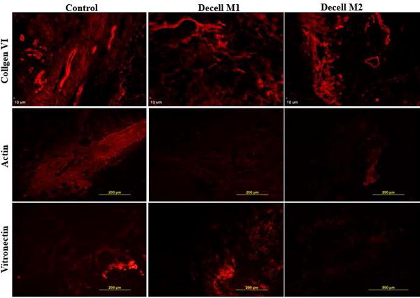
Figure 2.

Immunofluorescent staining for collagen VI, actin, and vitronectin. Paraffin fixed slides prepared with tissue from untreated controls or scaffolds decellularized using methods M1 and M2 were stained with fluorochrome labeled antibodies to collagen VI, actin, and vitronectin. Images are representative of n =3.
