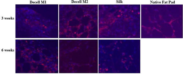
Figure 5.

Immunofluorescent Detection of CD 31. demonstrated the vascularization of the implanted scaffolds denoting their integration. Scaffolds were implanted into C57BL/6 mice transgenic for ubiquitous expression of the green fluorescent protein (GFP) for periods of 3 or 6 weeks. A silk scaffold implant and native adipose tissue served as positive controls. Sections were stained with DAPI for detection of nuclei (blue) or anti-CD31 fluorochrome labeled antibodies (red).
