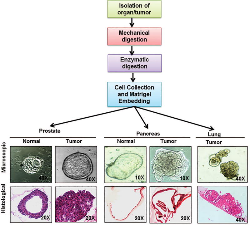
Fig. 2. General scheme of generating organoids and representative figures of organoids generated by our laboratory.

The flowchart represents the scheme of organoid isolation which is modified for each organoid according to the organ or tissue architecture to generate submerged organoids. Briefly, desired source tissue (progenitor cells or tissue fragments) is isolated from host by mincing the organ and then subjecting it to enzymatic digestion. The digestion media composition and the digestion protocol are decided depending on the host tissue. The digestion media usually contains a mixture or Dispase and Collagenase or Collagenase alone and can take from 30 mins to 4-6 hours. Following digestion, the cells are mixed in the matrix (like matrigel or collagen) suitable for the desired organoids. A suitable media is overlaid once the matrix solidifies. Once generated, organoids grow in ductal like morphologies like their human counter parts. Picture panels depict the microscopic pictures of organoids generated in our lab from normal and cancerous prostate and pancreas as well as lung cancer organoids (upper panel) along with hematoxylin and eosin stained sections of the same (lower panel) depicting the difference in organization of cells in each of these organoids. Figure magnifications are mentioned on each of the figures.
