Fig. 6.
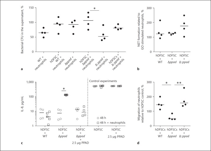
Interaction between P. gingivalis, hDFSCs and neutrophils in the triple culture. a Phagocytic activity of neutrophils. Analysis of the ability of neutrophils to phagocytose P. gingivalis strains in the presence of primed hDFSCs. The hDFSCs were incubated with WT, Δppad, or Δ::ppad for 24 h under anaerobic conditions. The bacterial control representing 100% is the amount of bacteria without neutrophils. b Analysis of the NET formation of neutrophils after 24 h of incubation with primed stem cells. Subsequently, the supernatants were sterile filtered, and freshly isolated neutrophils were incubated for 24 h under anaerobic conditions. Neutrophils incubated with untreated hDFSCs and activated with glucose oxidase for 2 h served as a control. Neutrophils activated by P. gingivalis strains were incubated for 2 h, and the formation of NETs was determined. The results were correlated with the glucose oxidase stimulation, which was defined as 100%. c The IL-8 concentration was measured in the culture supernatant. hDFSCs were primed with P. gingivalis WT, Δppad, or Δ::ppad under anaerobic conditions (open circles, compare with Fig. 2, Step I). After 24 h, neutrophils were co-cultured in sterile-filtered supernatants for another 24 h (open squares, compare with Figure 2, Step II). As a control, the IL-8 levels were quantified via ELISA after 48 h from the supernatant of the primed cells without neutrophils (open circles). Unprimed stem cells served as controls. d Analysis of neutrophil migration in a Transwell system. To this end, hDFSCs were primed with WT, Δppad or Δ::ppad. The supernatant was sterile filtered into the wells, and a suspension of freshly isolated neutrophils was added into the insert. The number of neutrophils in the wells was counted and compared with the medium control, which was defined as 100%. The results are displayed as the median; * p < 0.05, ** p < 0.01 were considered significant (ANOVA), n ≥ 4 (each data point represents 1 independent biological replicate).

